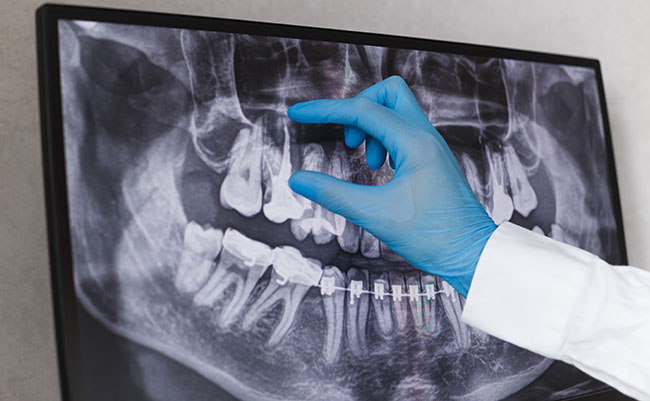
A standard procedure that is part of most annual dental appointments is taking dental x-rays in order to see bone levels for each tooth and tooth positioning, as well as to monitor the development of teeth in young children and teenagers. X-rays are taken in a complete oral exam. They can also show areas of infection, dental decay (cavities), and tartar deposits.
Diagnostic dental x-rays (dental radiographs) play a key role in oral health. They help us visualize diseases of the teeth and surrounding tissue that cannot be seen with a simple oral exam, and enable us to review your oral health development, as well as identify underlying problems related to the teeth, jaw and soft tissues of the mouth.
Dental x-rays show a full tooth picture from the top of the tooth to the very tip of the root. It is necessary to see your teeth below the gumline and detect potential tumors, oral cysts, tooth decay, and dental bone loss.
Not everyone needs to get yearly x-rays, especially those who have no apparent dental problems. The American Dental Association (ADA) reports that adults who brush properly and take good care of their teeth (and have no cavities or gum/oral conditions) only need X-rays every couple of years, and up to every three years.
While safe, dental x-ray do require very low levels of radiation exposure. The risk of potentially harmful effects very small.

